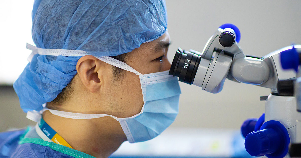
A View Into the Importance of Annual Eye Exams
How routine ophthalmologic examinations can provide the first indication of a serious health problem
Reviewed by: Gene Kim, MD
Written by: Erich Pelletier

The annual eye exam - millions of people make this appointment to assess overall eye health, to update optical prescriptions, and to check for signs of problematic conditions, such as cataracts and glaucoma. But routine ophthalmologic examinations can also provide the first indication of serious health problems well beyond the eyes.

We spoke with board-certified ophthalmologist Gene Kim, MD, a corneal transplant specialist and anterior segment reconstructive surgeon in UT Health Austin’s Mitchel and Shannon Wong Eye Institute, about how the eyes can be used to detect and assess systemic health issues, what “20/20 vision” really means, why dilating the eyes is not always needed during an exam, and one of the most common behaviors putting people’s vision at risk.
Why are annual eye exams important in ways that go beyond vision?
If a patient is between the ages of 18 and 64 and has no known or suspected vision problems, ophthalmologists recommend scheduling a complete eye examination (beyond a vision test) at least once every two years. Patients 65 years of age or older with no known or suspected vision issues should schedule their eye exams on an annual basis, a least once every year. Adults with known or suspected vision problems should schedule, at minimum, a yearly exam regardless of their age.
Nearly half of Americans between the ages of 25 and 38 don’t believe they need an eye exam if their vision is clear. However, ophthalmologists can identify early warning signs and other manifestations of more than 270 chronic systemic diseases through a standard eye examination.
The rear interior surface of the eye is called the retina, a layer of neural tissue that senses light and, through the optic nerve, sends signals the brain interprets as images. The retina is packed with blood vessels, nerves, and connective tissues. These structures can move, break, degrade, or otherwise change as a result of problems elsewhere in the body, providing a set of indirect signals about a patient’s overall health.
Dr. Kim likens the retina’s blood vessels to plumbing. “What’s going on within the pipes in the eyes is going on elsewhere in the body. And so, I always want to ask, ‘Are the pipes good? Are they damaged? Are they leaking? Are they broken?’ Because when the pipes are damaged in the eye, there’s likely a cause that can be found somewhere else in the body.”
Other aspects of the eyes are also tied to a patient’s overall health. Pressure levels within the eyes, coloration of the eye’s lens or cornea, inflammations in various parts of the eyes, or issues with the muscles surrounding and controlling eye movements can also signal more generalized disease.
“If you have inflammation in the body, a lot of times, your eyes’ blood vessels will leak, too,” explains Dr. Kim. “There are a lot of systemic diseases that actually manifest themselves in the eyes. It’s a window into what is going on inside the body.”
Major conditions that can be detected and assessed through eye examinations include:
- Brain tumor: Elevated pressure in the brain caused by a tumor can be transmitted to the eyes. Changes to the optic nerve, to the pupils, and to a patient’s vision can all be signs of tumors in the brain.
- Diabetes: High levels of blood sugar can cause diabetic retinopathy in which blood vessels in the retina swell and leak, degrading vision substantially. At times, retinopathy is the first indication of diabetes.
- Heart disease: Strokes can occur in the retina’s blood vessels due to heart disease. These strokes leave behind marks on the retina that may be detected by ophthalmologists.
- High cholesterol: Discolorations in a ring shape around the cornea, as well as other issues with retinal blood vessels, can indicate high cholesterol levels in a patient.
- Multiple sclerosis (MS): Inflammation of the optic nerve can be a sign of MS, a degenerative disease affecting the nervous system.
Numerous other diseases, ranging from high blood pressure to sexually transmitted infections, can also be detected through eye examinations.
Is it always necessary to dilate the eyes during a routine eye exam?
Most eye patients are familiar with the process of dilation, when drops are introduced into the eyes causing the pupil (the small black dot in the center of the iris, or the colored part of the eye) to widen. Similar to drawing back the curtains on a window, dilation allows ophthalmologists and technicians to see more clearly inside the eye when diagnosing potential problems. For many patients, pupillary dilation is a standard part of an annual eye exam.
“To be honest, patients hate being dilated,” says Dr. Kim. “Their vision is blurry for six to eight hours, and they can’t function properly.” Waiting 20 to 30 minutes for the pupils to fully dilate also adds to the length of the exam.
But technology can help. “Our technology has advanced quite a bit,” continues Dr Kim. “We have a wide field camera that can pretty much capture everything you see on a dilated exam without being dilated. It’s literally a photo. A lot of our technicians can do it, and I have migrated toward using this technology in my practice at the Mitchel and Shannon Wong Eye Institute instead of dilation.”
The advantages provided by digital imaging of the eye’s interior are numerous. “Capturing that photo allows us to complete the full exam in 15 to 20 minutes as opposed to the 45 minutes to an hour it takes with dilation,” explains Dr. Kim. “And it’s beneficial in the long run. It provides a library of the patient’s images that I can pull up and lay out to go back and look at images from previous exams.”
“I use the photos as a screening tool,” continues Dr. Kim. “If I see something abnormal, then I’ll dilate, as this is still the standard of care.”
What does “20/20 vision” really mean?
You have likely heard the term “20/20” to describe “perfect” vision. But what does the terminology actually mean?
“It’s all comparative to the ‘ideal’ person’s vision and what they can see at 20 feet, or whatever distance unit might be used,” explains Dr. Kim. “If you’re 20/20 and you’re being compared to that ideal person, then what they can see at 20 feet, you can also see at 20 feet.”
“But if you’re 20/40,” he continues, “what the ideal person sees at 40 feet is what you would be seeing at 20 feet. And you can go further and further. With 20/200 vision, an object seen clearly by someone with perfect vision at 200 feet away would not be seen clearly by you unless you were only 20 feet away. So, it’s all comparative based on what the standardized, ‘perfect vision’ person can see.”
What are some of the best practices patients can follow outside of scheduling routine eye exams?
“I think one of the most important things for people to be careful of are contact lenses,” says Dr. Kim. “I’m a cornea specialist, and a third of the major eye problems that I see – one of the leading causes of complications, such as needing a corneal transplant – is from horrific infections caused by contacts.”
Prescription contacts are better. “They’re very breathable, very smooth,” continues Dr. Kim. “But there are a lot of contacts out there that are horrendous, especially those that are sold for cosmetic purposes or for Halloween. Basically, they can suffocate your eyeball, and they can rub the cornea. Sadly, we see a lot of young college students who come in with terrible eye infections, which can lead to permanent scarring that sometimes requires a transplant.”
Even high-quality contact lenses can cause issues if not properly maintained. “A lot of times, people don’t care for them,” explains Dr. Kim. “They become like that dirty sponge you have by your sink, holding all this infection. People go to the pool, and all that gross water is soaked up and held against the eye like in a plastic bag. I’ve done plenty of corneal transplants for people who have misused their contacts and have had holes in their eyes from infection.”
“If there’s one thing that I could communicate to everyone, it’s that contacts are super dangerous,” says Dr. Kim. “I think people don’t even realize how dangerous they actually are.”
More than 45 million Americans wear contact lenses. Are you one of the 40-90% of Americans who do not properly follow contact lens care and safety instructions? Explore the contact lens safety tips below.
Contact lens safety tips:
- Do not sleep in your contact lenses
- Wash your hands with soap and water (and dry them with a clean cloth) before handling your contact lenses
- Keep your contact lenses away from all water
- Clean your contact lenses with the proper contact solution
- Do not wear your contact lenses past their expiration date
To make an appointment with UT Health Austin’s Mitchel and Shannon Wong Eye Institute, call 1-833-UT-CARES (1-833-882-2737) or visit here.