You Can’t Take a Picture of Pain
Why the most important part of treating lower back pain is the connection between a patient and a clinician
Reviewed by: Mark Queralt, MD

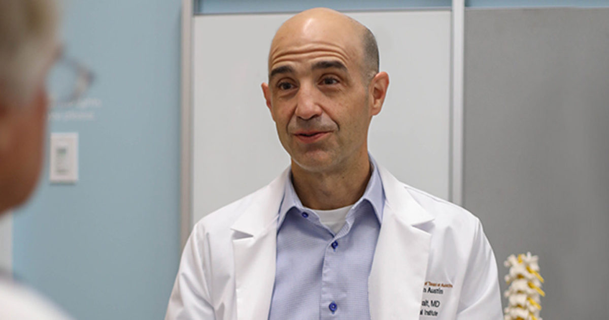
In recent decades, amazing advances in medical imaging have transformed health care. While today’s X-Rays, MRIs, PET scanners and other imaging systems can non-invasively capture ever-more detailed images of the deepest of the body’s internal structures, as board-certified physical medicine and rehabilitation specialist Mark Queralt, MD, Clinical Director of the Back and Neck Pain Center within UT Health Austin’s Musculoskeletal Institute observes, to date, there is no imaging system that can take a picture of pain.
“It’s estimated that as many as 80 percent of adults will experience lower back pain during their lifetime; and back pain is the leading cause of disability for people around the world,” says Dr. Queralt. People miss work; their lives are disrupted; they can’t sleep—it can be a really serious problem.”
Learn more about the relationship between sleep and pain.
“When an X-Ray or an MRI shows a bulging disk, or a bone with signs of age-related wear and tear, it can be really tempting for patients believe that doing something to the physical structure will make the pain go away,” notes Dr. Queralt.
“It’s important to remember that physical changes over time are extremely common, to the point that 96 percent of people over the age of 80 have some level of disk degeneration,” continues Dr. Queralt. More often than not, these ‘abnormalities’ are asymptomatic—which means that they don’t always cause pain.”
Additionally, imaging may not tell the whole story. “An estimated five to 50 percent of patients report unsuccessful outcome following lumbar spine surgery (commonly called failed back surgery syndrome),” warns Dr. Queralt. “It is very important to understand the significance of imaging findings before undergoing aggressive intervention.”
As such, images are just one of the three important tools that the Back and Neck Pain Center uses when caring for patients. “The evaluation of back pain is a process that involves a collaboration of medical and rehabilitation experts,” says Dr. Queralt.
The Back and Neck Pain Center begins the evaluation with a thorough review of a patient’s history:
- When did the pain begin?
- What makes it worse? What makes it better?
- Are there any other issues involved that might indicate that the pain is part of a larger set of problems?
Next, patients undergo a thorough physical examination during which the spine team compares the patient’s descriptions of their pain and function to the results of their exam (which must include maneuvers to assess response to stressing and de-stressing potentially painful structures).
The third part of the process, if needed, may be a set of images of the area where the pain occurs. The combination of a patient’s history and the clinical care team’s observations creates a detailed picture of that patient’s unique and personal circumstances.
“Because many of the degenerative changes that can appear on an imaging study are a natural part of the aging process, and often don’t result in pain, it’s very important that patients and clinicians work together to create a full picture of a person’s situation,” concludes Dr. Queralt.
“Chronic pain is a complex and personal experience. Addressing it effectively requires a purposeful diagnostic method and a treatment plan that is the right mix of physical rehabilitation, medications, and other approaches leading up to and, in some appropriate cases, including surgery,” says Dr. Queralt. “While the goal of this process is always to arrive at a treatment approach that is uniquely right for you, the first step, in every case, is connecting with your own, personal care team.”
For more information about the Back and Neck Pain Center or to request an appointment, call 1-833-UT-CARES or visit here.