Are All Forms of Glaucoma Alike?
A fellowship-trained glaucoma specialist from UTHA discusses the different types of glaucoma
Reviewed by: Eileen Bowden, MD
Written by: Lauren Schneider

As the second-leading cause of blindness, glaucoma affects millions of people worldwide. Within this population, individuals may experience drastically different symptoms, as glaucoma is an umbrella term for several conditions.
“Glaucoma is a group of diseases that all damage the optic nerve, the cable that connects the eye and the brain,” explains Eileen Bowden, MD, a fellowship-trained glaucoma specialist in UT Health Austin’s Mitchel and Shannon Wong Eye Institute.
The optic nerve is responsible for transmitting visual information to your bran. Damage to the optic nerve is typically a result of elevated intraocular pressure, which is caused by the buildup of fluid inside the eye. “You can think of this increased pressure as squeezing on the optic nerve, often causing irreversible damage to the nerve.” Although not all types of glaucoma are associated with elevated intraocular pressure, many forms of glaucoma are distinguished by what causes intraocular pressure to rise.
Explore a glaucoma Q&A with Dr. Bowden.
<br>Types of Glaucoma
Glaucoma is classified by whether it is caused by another medical condition (secondary glaucoma) or it has no known cause (primary glaucoma).
Primary glaucoma
Primary glaucoma is not associated with an underlying medical condition.
Types of primary glaucoma include:
- Open-angle glaucoma: This is the most common form of glaucoma in which symptoms of vision loss occur gradually until the individual develops tunnel vision. While many cases of open-angle glaucoma are caused by high intraocular pressure, individuals with normal eye pressure can also develop this condition. Researchers are still working to determine the exact cause of open-angle glaucoma.
- Closed-angle glaucoma: Under normal conditions, intraocular pressure is maintained by the eye’s constant production and drainage of fluid. Fluid is drained from a portion of the front of the eye known as the drainage angle. In closed-angle glaucoma, also known as angle-closure glaucoma, the iris (colorful portion of the eye) bulges forward, blocking the eye’s drainage canals. This results in fluid buildup in the eye and leads to increased intraocular pressure, which can occur gradually or very quickly. When a rapid buildup of fluid within the eye is accompanied by a sudden onset of symptoms, this is referred to as acute closed-angle glaucoma. This condition can be painful and is often accompanied by blurry vision, headache, and nausea. Because this form of glaucoma progresses so quickly, it should be treated as soon as possible to avoid permanent vision loss.
Secondary Glaucoma
Secondary glaucoma is caused by an underlying medical condition.
Types of secondary glaucoma include:
- Traumatic Glaucoma: This form of glaucoma develops following traumatic injury to the eye or head. Both penetrating injuries from sharp objects and blunt trauma can cause traumatic glaucoma.
- Neovascular Glaucoma: Individuals with diabetes often develop abnormalities with their blood vessels, including those in their eyes. These blood vessels can grow over the eye’s drainage system and block fluid from leaving the eye. This results in increased intraocular pressure and optic nerve damage. Neovascular glaucoma should be treated with urgency due to the condition’s rapid onset. Neovascular glaucoma is not the only ophthalmologic condition associated with diabetes. Find out more about diabetic retinopathy and why it’s a common complication of diabetes.
Congenital glaucoma
Glaucoma can be present from infancy in children who are born with a condition that prevents the drainage of fluid from the eye. Children with congenital glaucoma are often described as having large cloudy eyes that are sensitive to light. Some forms of congenital glaucoma are primary, while others are secondary glaucoma because of their association with some other health condition. Regardless of cause, congenital glaucoma needs to be treated quickly to preserve the baby’s vision.
Treating Glaucoma
“Types of glaucoma that develop quickly, such as acute closed-angle glaucoma or neovascular glaucoma, must also be treated quickly,” warns Dr. Bowden. “These forms of glaucoma are more debilitating than the slower-moving types of glaucoma that allow us additional time to figure out what’s going on and come up with a personalized treatment plan.”
While certain types of glaucoma require more immediate care, treatment often involves many of the same interventions regardless of the cause. First, eye drops and oral medications are used to reduce intraocular pressure. If additional intervention is needed, laser treatment or surgery is performed to enhance the eyes’ drainage system. For more aggressive forms of glaucoma, laser treatment and/or surgery may be employed sooner.
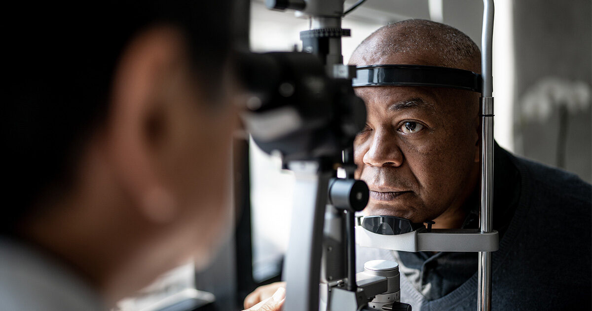
Glaucoma treatment is most effective when undertaken before disease progression causes permanent damage. “Recent technological advances have made this easier than ever,” explains Dr. Bowden. “We have a lot of new ways to test for glaucoma that didn’t exist fifteen to twenty years ago, allowing us to catching the condition earlier and earlier.”
Diagnosing Glaucoma
Early diagnosis can help preserve vision and prevent permanent vision loss. Glaucoma can be diagnosed during a comprehensive eye exam. According to the American Academy of Ophthalmology’s official guidance, adults should undergo such screening at least once by the age of 40. However, recommended exam frequency is based on an individual’s age and risk of glaucoma.
Adults with risk factors, such as family history or prior traumatic injury:
- Under 40: Exam every 2-4 years
- 40-54: Exam every 1-3 years
- 55+: Exam every 1-2 years
Adults with no known risk factors:
- One exam between ages 20 and 30
- Two exams between ages 30 and 40
- 40-54: Exam every 2-4 years
- 55-64: Exam every 1-3 years
- 65+: Exam every 1-2 years
For more information about the Mitchel and Shannon Wong Eye Institute or to schedule an appointment, visit here or call 1-833-UT-CARES (1-833-882-2737).