Breast Density Matters
FDA requires all mammography centers to notify women about their breast density after undergoing a screening mammogram
Sourced from: American Cancer Society, Centers for Disease Control and Prevention (CDC), and National Institutes of Health (NIH)
Written by: Ashley Lawrence

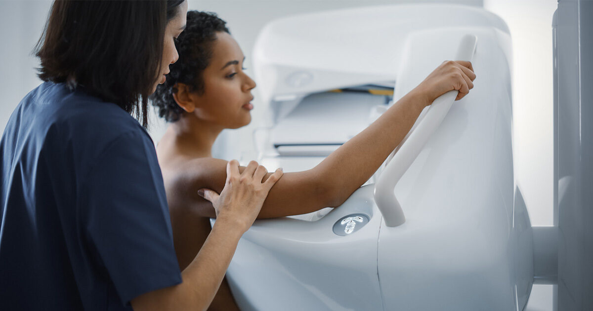
On March 9 2023, the U.S. Food and Drug Administration (FDA) published updates to mammography recommendations that now require all mammography centers in the U.S. to notify patients about their breast density after undergoing a screening mammogram. Screening mammogram reports sent to patients must now include information regarding the density of the patient’s breasts, which should be described as either “dense” or “not dense.”
What is breast density?
Breast density is a measure used to describe mammogram images. It is not a measure of how the breasts feel. Breasts are made up of glandular tissue (responsible for milk production), fibrous connective tissue (provides structural support), and fatty tissue (contributes to overall size and appearance). Dense breast tissue has relatively high amounts of glandular tissue and fibrous connective tissue and relatively low amounts of fatty breast tissue.
There are four categories of breast density:
- Category A: Fatty breast - Breasts are almost all fatty tissue.
- Category B: Scattered fibroglandular density - There are scattered areas of dense glandular tissue and fibrous connective tissue.
- Category C: Heterogeneously dense - More than half of the breast is made up of dense glandular tissue and fibrous connective tissue.
- Category D: Extremely dense - Most of the breast is made up of dense glandular tissue and fibrous connective tissue.
Breasts are classified as “dense” if they fall in the heterogeneously dense (C) or extremely dense (D) categories.
How do I know if I have dense breast tissue?
Nearly half of all women 40 years of age and older have dense breast tissue. Breast density can only be identified after a screening mammogram has been conducted and cannot be determined through self-examination. Once a screening mammogram has taken place, a radiologist will evaluate the mammogram results and categorize the breast density accordingly.
Does having dense breast tissue increase my risk of developing breast cancer?
Women who have dense breast tissue have a higher risk of developing breast cancer than women with less dense breast tissue. It’s unclear at this time why dense breast tissue is linked to breast cancer risk. It may be that dense breast tissue has more cells that can develop into abnormal cells.
Dense breast tissue can also make it challenging for radiologists to detect abnormalities on a screening mammogram. Whereas fatty tissue appears almost black on a screening mammogram, dense breast tissue appears white, making abnormalities such as breast masses and cancers, which also appear white, more difficult to identify.
If I have dense breasts, do I still need to schedule regular screening mammograms?
Most breast cancers can be seen on a screening mammogram even in women who have dense breast tissue, so it’s still important to schedule regular mammograms. Individuals at average risk for developing breast cancer are recommended to get screening mammograms every two years beginning at age 40. If you have a strong family history of breast cancer involving an immediate family member, such as a mother or sister, it is recommended that you begin scheduling screening mammograms when you reach 10 years younger than the age at which they were diagnosed or by the time you reach the age of 40, whichever comes first.
Do I need to take additional precautions if I have dense breast tissue?
While there are no specific recommendations or screening guidelines for women with dense breasts, 3D screening mammograms and breast ultrasounds can better detect abnormalities in dense breast tissue. Your healthcare provider may suggest other types of breast imaging in addition to regular screening mammograms.
To schedule a screening mammogram through UT Health Austin’s Imaging Center, please call 1-512-495-5231 or visit here.